
.
Ultrasonography works by sending a beam of sound waves into the body. When the beam hits the tissues, it is partly reflected back towards the probe. The probe detects the return signal and the machine processes this to produce a picture.
Different tissues reflect different proportions of the beam, and so produce different shades of white or grey in the ultrasound image. For example, bone reflects all of the ultrasound beam and so looks bright white on the screen. Tendons reflect some of the beam and appear grey. Fluid doesn’t reflect any of the beam and so looks black. This allows us to identify different structures and changes in the appearance of the structures we examine and guide our diagnosis. Like bone, air reflects all of the ultrasound beam and prevents us from seeing any structures behind it. This is why we need to clip and apply coupling gel to the skin for scanning – clipping removes the air trapped between the coat’s hairs, and the coupling gel ensures the probe has an even contact with the area we want to scan.
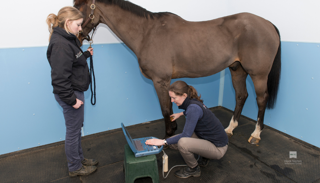
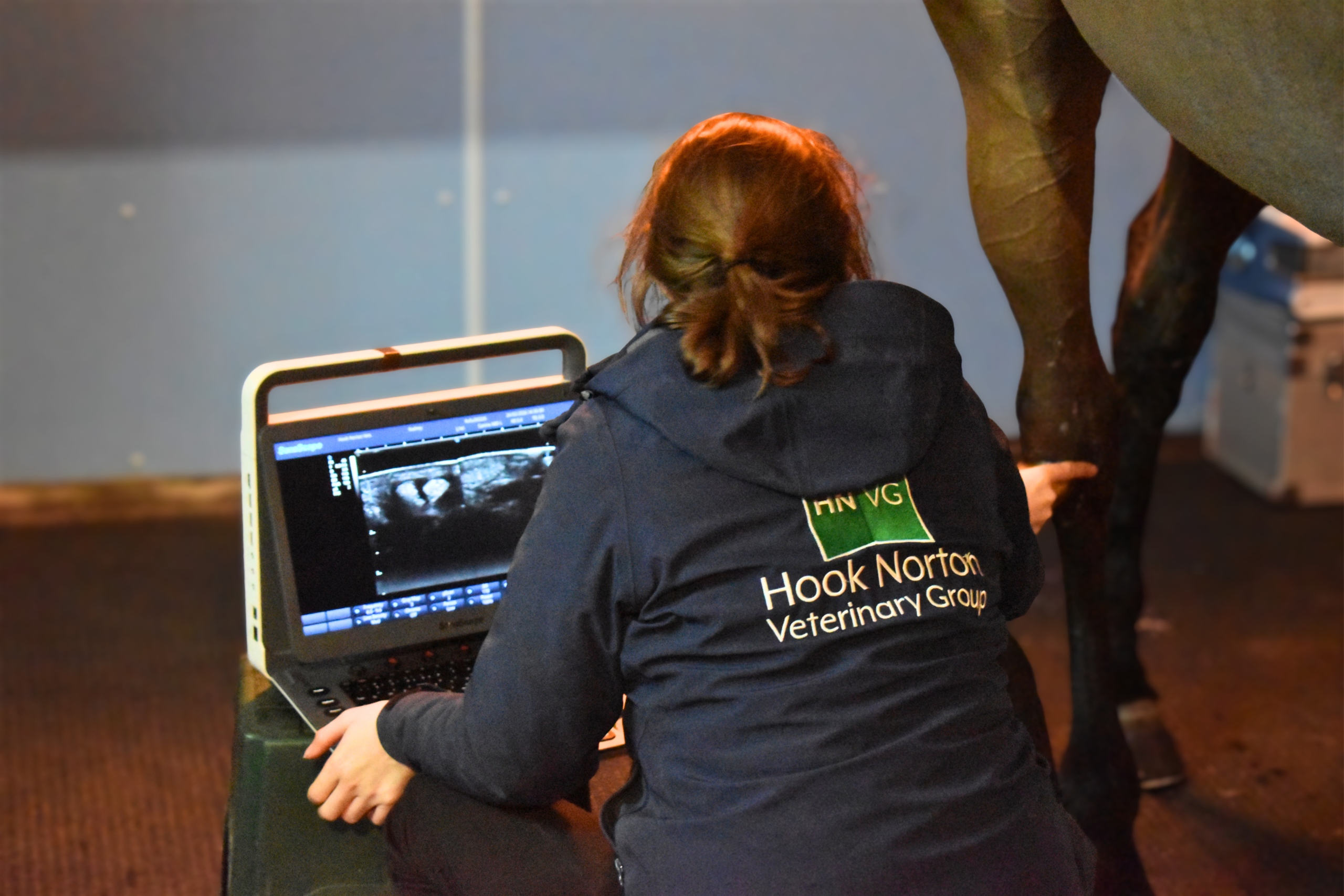
Scans of the lower legs are very commonly performed to diagnose and assess the severity of an injury. This information helps us to form a treatment plan tailored to your horse. In addition, repeating scans through the rehabilitation period lets us monitor healing and make sure that rehabilitation is progressing at the correct rate. Ultrasound scans of the tendons during the competition season are also a useful tool for monitoring horses performing at a very high level, such as eventers and racehorses, and can allow minor strains to be detected and treated before they become serious injuries.
We can also use ultrasound to examine joints, for example, the cartilages in the stifle joint or small OCD fragments in fetlocks and stifles. In these cases, ultrasonography complements the information gained from x-raying.
Ultrasound can be used to help us assess the abdomen in sick horses. The depth of tissue and the presence of gas in the large intestine means that not all of the abdominal contents can be seen on a scan, but there is still a lot of information that can be gained from imaging the structures we can reach! Ultrasound can help guide us in taking biopsies in horses with liver disease and in finding bladder stones.
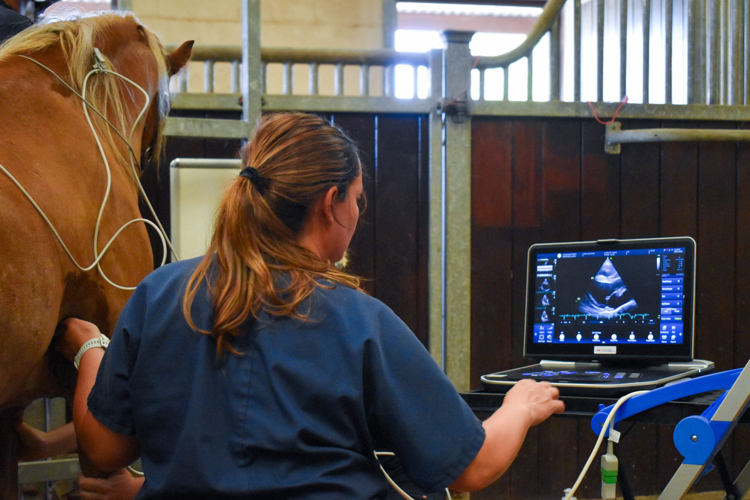
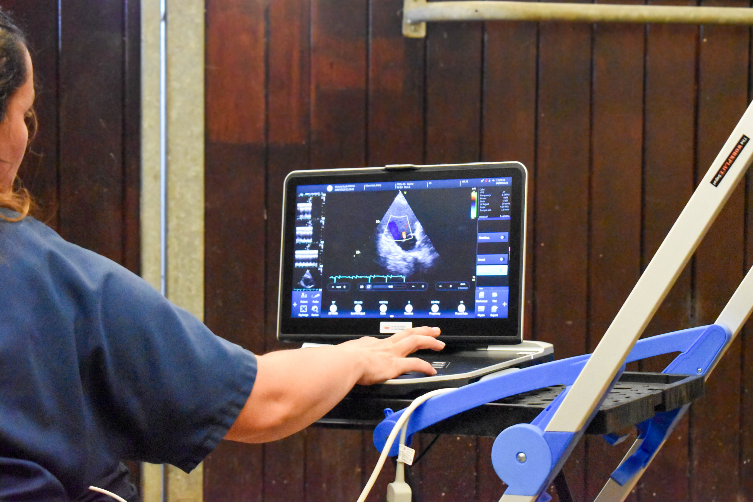
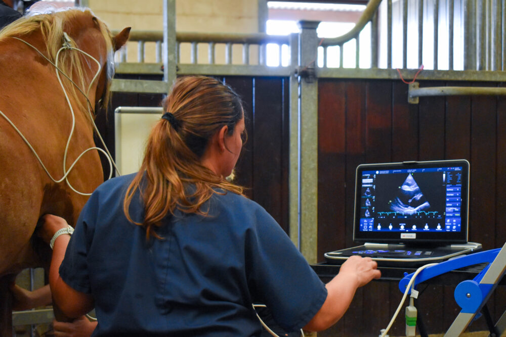
Cardiac scans (echocardiography) allow us to assess the health and function of the heart – our Doppler probe lets us identify turbulent blood flow caused by leaky heart valves.
Most of the time, we can examine the back of the eye using an ophthalmoscope, but if the cornea is completely clouded over, we can use ultrasound to check for problems such as retinal detachment and glaucoma.
Ultrasound examination can detect pregnancy as early as 14 days and find the embryo’s heartbeat from 28 days. It also lets us check the health of the placenta. View our Stud Work page for more information on getting your mare in foal.
Although bone reflects ultrasound, fractures of flat bones such as the pelvis and shoulder blade can sometimes be seen on an ultrasound scan. In a normal horse, the surface of the bone appears as a smooth, continuous white line. However, a fracture may create a gap or step in the white line – sometimes, this can be very subtle and may require scanning on several occasions to be sure of the diagnosis.
If you’re worried about your horse and would like to speak to one of our vets then please call your nearest branch or send an email.
Hook Norton: 01608 730085
equine@hooknortonvets.co.uk

